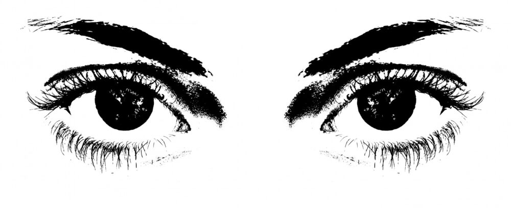
Anyone would love savoring a scoop of refreshing ice cream on a scorching summer day or cozying up with hot chocolate during chilly winters. However, life is anything but simple for diabetics. It is a complex journey with twists and turns that can impact vital organs like the heart, kidneys, nerves, blood vessels, and even the eyes, introducing a host of complications. A connection between diabetes and eyes might sound unusual, but the condition surprisingly does exist, called Diabetic Retinopathy (DR).
Diagnosing DR poses challenges as it typically lacks symptoms early on; however, it advances through the following stages:
Stage 1: Mild non-proliferative DR (NPDR), which is characterized by the presence of small red dots scattered across the retina, referred to as microaneurysms.
Stage 2: Moderate NPDR, which is marked by the emergence of cotton wool spots and dot hemorrhages, which look like little red spots.
Stage 3: Severe NPDR, which displays an appearance of venous beading and blood vessel abnormalities inside the retina.
Stage 4: Proliferative DR (PDR), which is characterized by the growth of new blood vessels (neovascularization) within the retinas. These are fragile and bleed. Minimal bleeding results in the appearance of floaters, while extensive bleeding leads to vision loss.
High blood sugar, or hyperglycemia, harms the retina’s blood vessels, causing DR. Hyperglycemia activates the polyol pathway, converting glucose into sorbitol, which accumulates in retinal blood vessels, causing microaneurysms and pericyte (eye cells) loss. Certain protein pathways are activated, leading to loss of pericytes and endothelial cells (blood vessel cells) and disruption of the blood-retinal barrier, which eventually results in retinal swelling. To compensate, the immune system produces growth factors that result in abnormal vessels, which are prone to rupture and risk vision loss. Regular eye check-ups are crucial for diabetics.
The eye in AI
In the healthcare sector, there is a growing anticipation of increased utilization of AI and deep learning algorithms in the next decade. Within ophthalmology, AI has already found practical applications in diagnosing DR by analyzing color fundus photographs, which are retinal pictures. Research has demonstrated the remarkable accuracy of AI systems in detecting DR. There are currently two AI systems in active clinical use in the United States: IDx-DR, the first FDA-approved AI algorithm for DR diagnosis that is usable by non-ophthalmic healthcare professionals; and EyeArt, the first FDA-cleared AI technology for autonomous DR detection. Additionally, Singapore Eye Lesions Analyzer (SELENA+), which employs a deep learning system, has gained regulatory approvals from multiple health organizations around the world.
Economically, integrating AI into large-scale DR screening programs could save costs. However, certain challenges persist, including legal issues related to diagnosing other medical conditions and the potential compromise in the accuracy of AI algorithms due to low-quality images. Despite these challenges, diverse AI applications in DR screening offer improved disease prediction and treatment guidance.
Print your vision
Replicating human retinas using 3D printing might sound unrealistic, but it is not impossible. Scientists have successfully developed retinal organoids, which come from a type of human cell called induced pluripotent stem cells. These organoids are made from a person’s cells, which means the treatment can be personalized to their needs. These organoids mimic real retinas and are crucial for studying eye disease treatments. Unlike previous methods using flat dishes or animals, organoids replicate human retinas more accurately, avoiding drug failures in human trials.
“These organoids are made from a person’s cells, which means the treatment can be personalized to their needs.”
While creating organoids is time-consuming and does not fully replicate late-onset eye diseases or a real retina’s complexity, scientists see their potential. Organoids can improve drug development, ensuring successful clinical trials by testing various reactions and checking for unintended side effects. Despite challenges, using retinal organoids is a promising advancement.
Although diabetes can bring challenges, there is a silver lining. Innovations such as AI diagnostics and retinal organoids are beacons of hope. They not only improve early detection and treatment, but they also lead to personalized and efficient therapies, safeguarding the invaluable gift of sight.
Related posts:
 Oral phenylephrine: placebo, in tablet form
 Ending autoimmune disease? ‘Inverse vaccination’ and unlearning immune response
 Bionic eyes: Can we provide artificial vision?
 Bite the bullet or face the fog?
 Crystal clear: Acrylic skull windows revolutionize post-craniectomy recovery
 Genetic interventions in Alzheimer’s disease: A promising future