It might sound like science fiction, but the future of 3D-printed organs is closer than one would imagine. New innovations in printing technology are making leaps in implants or grafts possible. One day, it might even be possible to replace major sections of the heart using cells from a patients’ own body.
One day, it might even be possible to replace major sections of the heart using cells from a patients’ own body.
3D printing has an important place in medical modeling and planning. Using volumetric imaging, a set of 2D photos can be “stacked” into a digital 3D model. Computer tomography, a scanning method that digitally combines multiple X-rays from different angles for a complete image, has been the main imaging technique. It provides submillimeter resolutions and can be safely used on people with implants.
From there, the type of printer and the materials depend on the purpose. Selective laser melting, which is used for medical implants, can create parts of metal or ceramic using a laser beam that melts the materials. More recently, the development of PolyJet technology, which jets thin layers of liquid photopolymers and hardens them with ultraviolet radiation, has become favored for patient-specific anatomical models.
Patient-specific models can be critically important in many ways.
Patient-specific models can be critically important in many ways. They can be a tool for communicating with patients using color-coded or transparent polymers that help them understand their treatments. It can also help doctors with planning as models with accurate blood flow can be used to test prosthetics prior to using them on the patient. It can also help with planning for removal of calcifications, tumors, or other structural flaws.
Instead of printing with polymers, new work that uses a patients’ tissues to create a cardiac patch for defects is promising. From the tissue, induced pluripotent stem cells (iPSCs) and hydrogel can be created. Using the hydrogel as a structural guide, the iPSCs can be used to print cardiac patches, complete with veins for oxygenation. The benefits of this technique are that the new cardiac patch is a complete match for the patient and that the body will not reject it.
While creating fully functional organs seems futuristic, a beating 3D-printed heart may not be far off.
The researchers also printed a small-scale human heart in a growth medium using human-derived stem cells and biological hydrogel. The heart was the first to be created with fully functional contraction mechanisms. Although it is only about the size of a rabbit’s heart and cannot pump itself, it is an important step to possibly creating organs complete with all of their features. While creating fully functional organs seems futuristic, a beating 3D-printed heart may not be far off.
Image Source: UCSD Jacobs School of Engineering
Related posts:
 Fixing a Broken Heart: Treatment of Cardiac Valve Disease
 Keep it FRESH: Printing the organs of the future
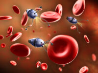 One nano step for man, one giant leap for mankind: Developments of nanotechnology and its applications in medicine
 Good vibrations (and how to sense the really bad ones)
 Pigs: The solution to organ shortages?
 Hidden Markov Models for biochemical applications