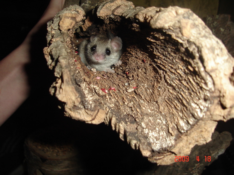
As the dreary winter winds arrive, animals previously roaming their environment during warm times are left with few options. Many animals choose to migrate, while others choose to hunker down for colder weather by hibernating. Most people understand hibernation to be a state when animals enter a deep sleep, preparing for this slumber by eating and storing enough food to last through the winter. However, this common perception has its foundations in biological and physiological changes.
Animals are extremely resilient creatures and can adapt to survive various conditions. Their method of protecting themselves from cold and food scarcity is by entering torpor, a physiological state where metabolic processes are suppressed to lower metabolic demand, conserve energy, and preserve body heat. During torpor in endotherms – warm-blooded animals – several cellular processes are blocked, resulting in low energy expenditure and thus significantly reduced body temperature.
Current data on torpor and the entrance into hibernation indicates that the process is orchestrated by the central nervous system; however, the physiological regulation of the process is not exactly clear.
Animals often naturally enter torpor in response to food shortages, of which hibernation is a primary example. However, during hibernation, animals must do more than gain a few extra pounds and prepare for the cold. According to a paper published by Nature, the metabolism, measured by rate of oxygen consumption, decreases notably in rodents during torpor, and their heart rates fall from 300 times per minute to almost undetectable levels. Since rodents can enter a hypometabolic state independent of cold weather in torpor, the neurological mechanisms orchestrating the process in rodents can reveal possible applications to humans.
This approach would be instrumental in medical techniques to slow tissue damage after strokes, especially because it inflicts no other damage to organs, tissue, and metabolic regulation.
A group of Japanese scientists with a variety of specialties such as cellular neurobiology, biosystems, and the brain studied laboratory mice to reveal how neuronal processes induce a torpor state. Current data on torpor and the entrance into hibernation indicates that the process is orchestrated by the central nervous system; however, the physiological regulation of the process is not exactly clear. This group of scientists is one of many questioning how this discovery can be applied to biomedical science, including superior treatments for stroke and brain trauma patients.
Although wild mice enter torpor during hibernation, laboratory animals do not experience this phenomenon as a result of their living conditions. Nevertheless, the laboratory mice studied occasionally displayed instances of short term torpor. The scientists targeted their research on the underlying neuronal circuit responsible for torpor in the hypothalamus, which is the region of the brain responsible for autonomous regulation such as body temperature, sleep, anxiety, and hunger. The group tested a series of drug injections at targeted coordinates in the hypothalamus to identify the neurons in the mice.
This science holds the potential for a novel treatment that could harness the protective benefits of torpor on the body to slow down cerebral inflammation incurred by strokes.
After robust testing of different drugs, they discovered that a protein called pyroglutamylated RFamide peptide (QRFP) was responsible for stimulating several hypothalamic neurons that trigger torpor. When QRFP is released in the hypothalamus, markers of torpor, such as body temperature and metabolic rate, confirm the identity and location of the responsible neurons. Although the mice in torpor remain in a hypothermic and hypometabolic state, their physiological systems remain functional, maintain the ability to self-regulate, and display no tissue damage post-spontaneous recovery.
Biomedical scientists are using discoveries surrounding medically induced torpor to make groundbreaking advances in the healthcare field.
The discovery of this neuronal mechanism could serve as a method to induce a hypometabolic state in non-hibernating species. This approach would be instrumental in medical techniques to slow tissue damage after strokes, especially because it inflicts no other damage to organs, tissue, and metabolic regulation. In the past, medical treatments have attempted to decrease metabolic rate and blood flow using therapeutic hypothermia to achieve similar results. However, this approach does not allow for the same thermoregulation and protection demonstrated in torpor. In a 2018 grant proposal, researchers from the University of Oxford began assessing the effects of torpor in mice such as its impactfulness on brain function and its effectiveness as a protective measure from brain damage. This science holds the potential for a novel treatment that could harness the protective benefits of torpor on the body to slow down cerebral inflammation incurred by strokes.
Biomedical scientists are using discoveries surrounding medically induced torpor to make groundbreaking advances in the healthcare field. By applying various studies of hibernation in rodents to humans, scientists aim to improve not only stroke treatment but also technology for post-heart attack tissue protection, organ preservation during transplantation, and diabetes. Additionally, as this research begins to gain recognition, NASA and other agencies are looking deeper into the future of this science for human space travel. The discoveries could be used to prevent muscle wasting during long distance space travel. As more research is targeted towards hibernating creatures, scientists are realizing the potential of applying this research to the realm of healthcare and beyond.
Nature (2020). DOI: 10.1038/s41586-020-2163-6
Related posts:
 Hibernation: When the past becomes the future
 The battle between hot and cold: How TRP channels are involved in temperature transduction in human skin
 Hot and touching research wins 2021 Nobel Prize
 Scientists on substances: Can researchers be open about drug use?
 Olive oil compounds as a potential therapy for Neurodegeneration
 Stop studying and go to bed: The impact of sleep on learning