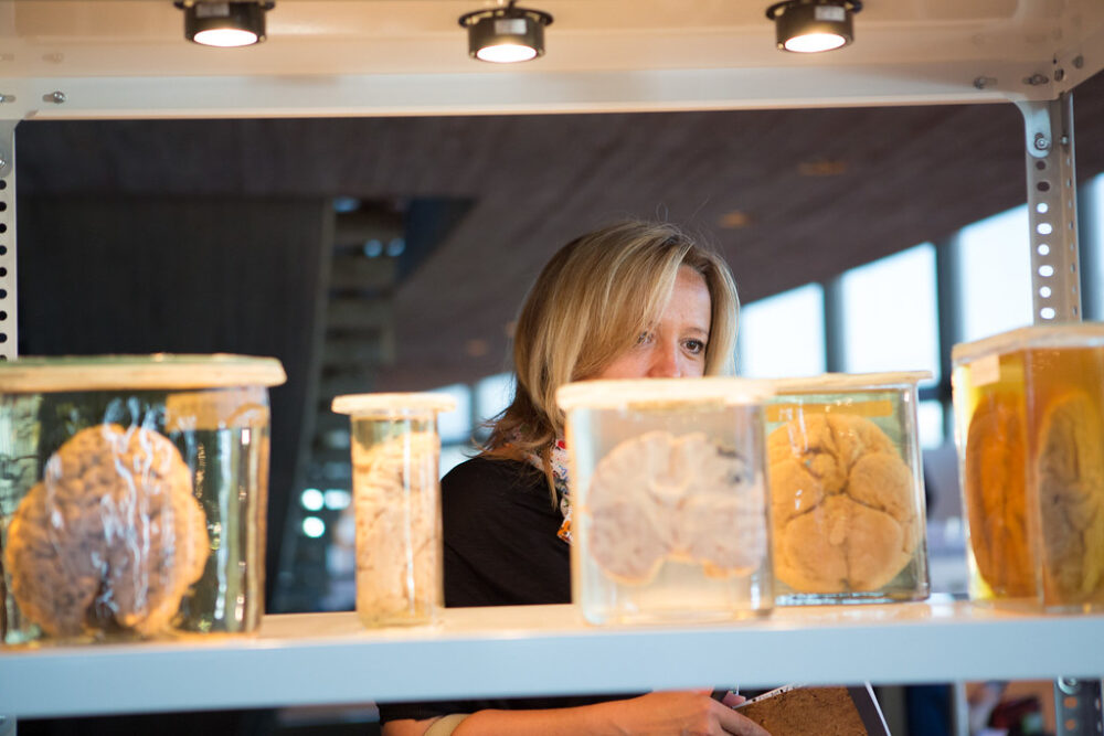
The immense capability of the human brain quickly disappears after death. A once–thriving organ built of intricate framework becomes pure mush, leaving behind an empty skull. Decomposition, marked by two processes, primarily affects soft tissues, the material from which organs are built. Within hours postmortem, autolysis causes the leakage of cellular enzymes into surrounding tissues, encouraging their breakdown. The brain is among the first to go — its high water content and intricate network of neurons and supporting cells make it difficult for the organ to remain intact even minutes after death. How is it then that some uncovered human remains include the fragile soft tissues of the human brain?
Scientists have explained that it is rare, but not necessarily unusual to find a preserved brain. A perfect storm of conditions could be the cause, but what’s puzzling is the numerous cases in which the brain is the only surviving soft tissue. Every soft tissue of the human body undergoes autolysis, from the liver to the white of the eye, and the brain is no exception. This provides an ideal environment for the proliferation of microbes, marking the beginning of putrefaction (a much kinder word for rotting). It should not be possible for a body, which underwent complete skeletonization thousands of years ago, to house a brain.
One of the most delicate soft tissues surviving conditions that the skin, liver, and heart couldn’t presents a bizarre phenomenon that can only be explained by the microanatomy of the brain itself. A skull uncovered in the U.K. in 2008, estimated to originate between 673 and 482 BCE, suggests that an ancient brain may not be as outlandish as it sounds.
The skull, excavated in Heslington, U.K., held a brain which was easily identified by its characteristic grooves. Upon antibody testing, it was found that aggregate, or clumped, proteins were present in high amounts. Proteins behave diversely depending on the order of their amino acids, strung together into long chains which can fold in on themselves. The clumped, tightly folded proteins in the Heslington brain consisted of neurofilaments and glial fibrillary acidic protein. Both filaments are described by researchers as “scaffolding” for the brain, and their behavior is likely the reason that the brain has survived millennia. The protein aggregates were present in high concentration in the outer grey matter of the brain, protecting the inner white matter from the process of autolysis. The ability of these proteins to fold tightly upon each other essentially sealed the brain off from the consequences of time, which ravaged the rest of the body. While autolysis beckoned in a microbiome and rapid decomposition of the rest of the body, the brain remained intact.
Interestingly, protein clumps like those found in the Heslington brain closely mirror the pathology of neurodegenerative diseases such as Alzheimer’s. Studying the behaviors of these ancient proteins creates a window into the still–living brains of 21st–century patients. By understanding how the protein aggregates degrade, an age–old brain could bring to life new conversations in modern medicine.


