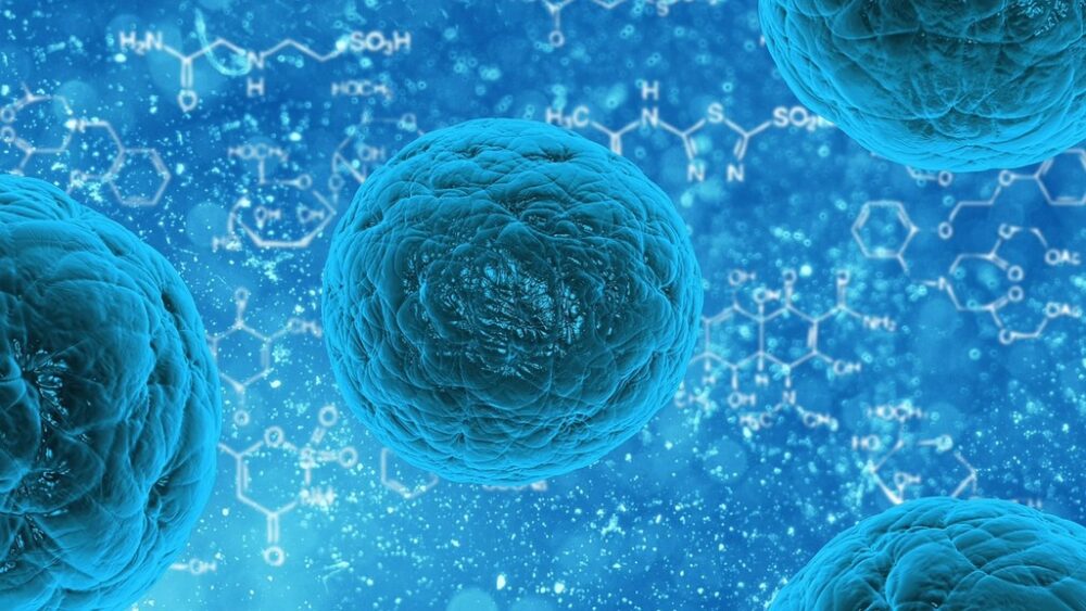
When you think of the skeletal system, what comes to mind? Most of us visualize a collection of rigid, porous bones, organized in “the leg bone’s connected to the knee bone” fashion. However, a significant portion of the skeletal system is actually composed of cartilage. In every anatomy textbook, you’ll find a section listing three types of cartilage: hyaline, elastic, and fibrocartilage. Hyaline cartilage, the most abundant type in the body, lines joints and envelops the ends of bones. Embryonic skeletons are composed primarily of hyaline cartilage and eventually ossify, or become bone. The growth plate, where long bones grow in length, is also hyaline cartilage. Tendons and ligaments are made of strong, dense fibrocartilage, and organs such as the larynx attain their flexibility and resilience from elastic cartilage.
In January 2025, a team of researchers at the University of California, Irvine upset the long-held belief that these three types of cartilage are the only classifications to exist. Their discovery of lipocartilage presents a potential new approach to regenerative medicine as well as a deeper understanding of unique lipid biology.
The finding describes cells that form and maintain stores of lipids which don’t change in the face of external factors, such as nutrient availability. Lipocartilage cells, or lipochondrocytes, were first reported in the mid-19th century by Dr. Franz Leydig, who described droplets of fat in rat ear cartilage. Until now, they’ve been largely ignored. Typical fat cells (adipocytes) are abundant underneath the skin and surrounding the organs, providing cushioning and energy storage. Their anatomy consists of one to several fat droplets, which range in size depending on the nutrients available. Conversely, no matter how limited the extracellular nutrients, lipochondrocytes maintain equally sized lipid vacuoles. Essentially, lipocartilage is a biological bubble wrap and provides a bouncy, supportive material from which tissues can be built.
Dr. Maksim Plikus of UC Irvine stated, “Currently, cartilage reconstruction often requires harvesting tissue from the patient’s rib – a painful and invasive procedure.”
Cartilage reconstruction is complicated because of the tissue’s lack of nerves and blood vessels. Because of this, chondrocytes, your average cartilage cells, replicate considerably slower than other cells of the body. Given the specific nature of chondrocytes, injuries to cartilage are oftentimes traumatic. Generating cartilage from a patient’s own tissue (by harvesting bone marrow stem cells) is frequently used for the treatment of joint injuries, offering pain relief and better mobility. In other cases, it is useful in treating birth defects or facial traumas. The significance of the rediscovery of lipocartilage lies in its stability and flexibility, thus making it an excellent candidate for techniques such as 3D printing tissues for patient-specific tissue engineering. Compared to standard cartilage generation practices, this presents an unexplored approach to a wide range of injuries.
The collaborative effort of scientists at UC Irvine and beyond showcases the relevance of reexamining centuries-old discoveries for a renewed approach to regenerative medicine. A focus on lipocartilage implicates new, versatile solutions to injuries which may otherwise be catastrophic. With the discovery at the Pilkus lab, it’s become clear that the way to bounce back from an injury may have been inside us all along.
Related posts:
 The unexpected ally: Can anti–seizure meds slow glioblastoma growth?
 It takes heart: Cardiovascular approaches to combat hypertensive disorders of pregnancy
 3D bioprinting: Ctrl+P human organs in real time
 Mind over medicine: The placebo effect
 Crawling cures: The potential value of insects in medicine