You may have heard of some eye-catching statistics like “every seven years, every cell in your body has been replaced” or “every day, 50 billion cells in the human body die,” and these are true. Cells within the body are constantly multiplying as you grow, as part of maintaining homeostasis. However, what you might not know is exactly how these cells are replaced. The vast majority of this cell death is, by design, occurring by a natural process termed apoptosis.
Apoptosis — also known as programmed cell death — is the process by which old or diseased cells are naturally killed to pave the way for new cells. This differs importantly from necrosis, which is cell death that results directly from injury or disease. Programmed cell death is just that — programmed. There are a few well-regulated and controlled steps along one of two pathways that must occur before the irreversible process is started.
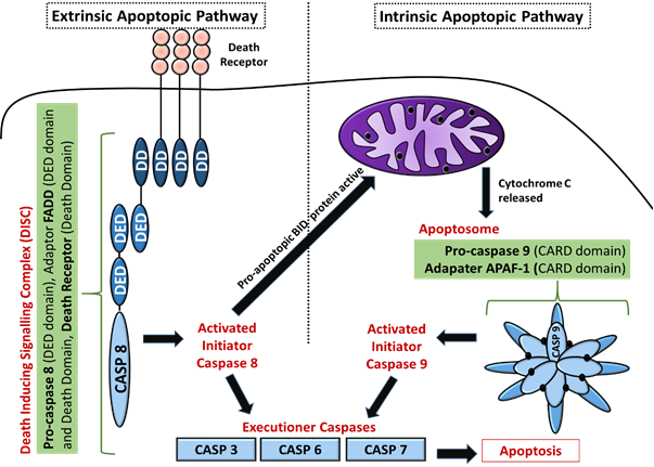
The first pathway is the intrinsic pathway. This is almost always instigated by some stressor, such as heat, radiation, hypoxia (lack of oxygen), or lack of nutrients. It can also be started by a high concentration of certain ions or molecules like calcium or fatty acids. If any of these conditions are sensed by regulatory proteins in the cell, they can send along a signal through a cascade of messenger proteins. This signal can be stopped if it is determined that the cell no longer needs to die (perhaps a cell previously undergoing hypoxia is now oxygenated). The message is sent toward the mitochondria, the essential, energy-producing organelles of the cell.
Once the mitochondrion is reached, the process of death begins. While apoptosis can be initiated by a number of various signals, the most common pathways all begin by increasing the permeability of the mitochondrial membrane.
In a healthy cell, the mitochondrial membrane maintains a gradient of protons on one side, which drive its energy-making capabilities. If the membrane is made more permeable, this gradient is lost, and the potential to create energy drops drastically. The mitochondrion will then begin to secrete molecules that speed along the apoptotic process. These molecules, called SMACs (second mitochondria-derived activator of caspases) recruit caspases, a certain class of enzymes that break down proteins, to digest much of the cell, leaving the rest to white blood cells. These enzymes break open the cell like a piñata, releasing its contents and leading to its demise.
The extrinsic pathway takes a slightly different approach. On the exterior of the cell membrane, there are two receptors that take part in inducing apoptosis: the TNF (tumor necrosis factor) and Fas receptors. Certain signaling proteins from elsewhere in the body can bind to these receptors, forming the death-inducing signaling complex (DISC). This complex then promotes the activity of caspases, which finish the job.
Despite apoptosis primarily being used to clear away dead or diseased cells, it is also a useful tool in both fetal development and overall growth. In the womb, the fingers and toes of a fetus grow first as a mass of cells. Careful apoptosis of certain cells allows each finger to be separated once they’re large enough. Apoptosis functions almost as a controlled demolition to pave the way for more refined growth.
While apoptosis is often used to promote growth and prevent the accumulation of weakened or dead cells, it can also be combated by tumors, leading to too many cells staying alive when they shouldn’t. Certain, carefully regulated processes within apoptosis can mutate and wreak havoc on the previously well-controlled system. In many cancers, inhibitors to various parts of the signaling cascades are overexpressed, preventing apoptosis from occurring. This allows the tumor to grow despite parts of the body signaling for it to be destroyed. It’s quite hard for the body to fight off a tumor when it literally can’t pull the trigger.
Despite cancer’s effective mechanisms of defense, there are some promising avenues of research within the apoptotic pathway. For example, researchers have been investigating creating their own apoptosis-inducing ligands to bind to the external TNF and Fas receptors to artificially create the DISC. Many times, the questions these scientists are studying boil down to how to enable cancerous cells to kill themselves, and at the end of the day, that’s all apoptosis is — it’s the basic mechanism of inducing death to allow for more life.
Image Source: Wikimedia Commons


